You are using an outdated browser. Please
upgrade your browser to improve your experience.
Volume 1 in the Series: The Johns Hopkins Atlases of Pathology

Johns Hopkins Atlas of Pancreatic Pathology
by Johns Hopkins Mobile medicine
What is it about?
Volume 1 in the Series: The Johns Hopkins Atlases of Pathology
App Screenshots

App Store Description
Volume 1 in the Series: The Johns Hopkins Atlases of Pathology
AUTHORS: Ralph H. Hruban, Bona Kim, Corrinne Sandone, and Toby C. Cornish
SERIES EDITORS: Toby C. Cornish, Norman J. Barker, and Ralph H. Hruban
This app is a teaching tool for medical students, residents, fellows, and practicing pathologists.
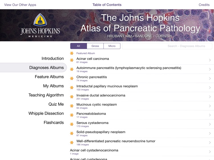
The Atlas of Pancreas Pathology is comprised of five modules: an interactive teaching algorithm, a searchable image atlas, an image-based quiz, flashcards, and a Whipple specimen dissection tutorial animation. Viewing multiple examples of the same entity or feature from this large, rich image atlas will strengthen your diagnostic skills.
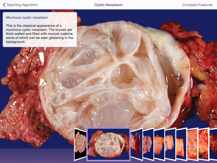
Teaching Algorithm: The teaching algorithm is a tool to teach the diagnostic criteria for the most common neoplasms of the pancreas. The algorithm consists of a series of usually dichotomous decision points, starting with determining if the tumor is solid or cystic, which end in specific diagnoses. Gross and microscopic photographs, together with didactic illustrations created by medical illustrator Bona Kim, support the instructional design of the algorithm. The algorithm was adapted with permission from RH Hruban, MB Pitman, and DS Klimstra, Tumors of the Pancreas. Washington D.C.: American registry of Pathology; 2007. Atlas of Tumor Pathology; 4th series, fascicle 6.
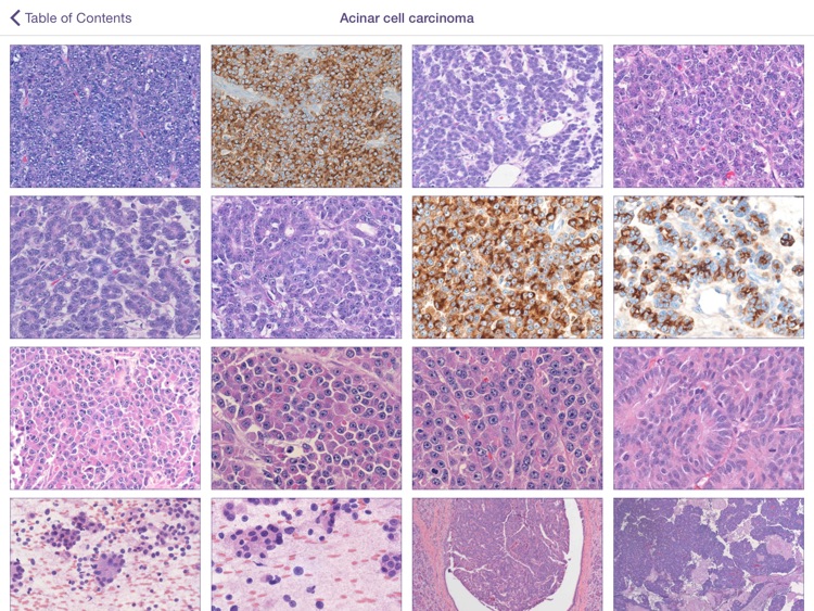
Image Atlas: The image atlas contains over 1,400 high resolution color images with captions authored by a leading expert, and covers 115 diagnostic entities. These gross and microscopic photographs can be viewed together or separately, and two entities can be compared, side by side. The atlas can be viewed by diagnosis or by features (these features re-enforce the diagnostic features used in the algorithm), and the atlas can be searched using a key word search. You can create your own albums and add images you select from from the atlas to these albums.
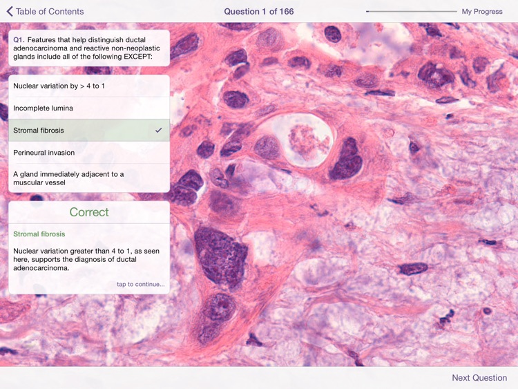
Quiz: The Quiz contains 166 multiple choice questions for self-study.
Flash Cards:The Flash cards module allows you to view a randomly generated assortment of images from the image bank and subsequently view the diagnosis and caption by simply “flipping” the card over.
Whipple Dissection: This instructional video, expertly illustrated by Anastasia Demson, illustrates our approach to the gross dissection of a Whipple specimen.
Explore this app and improve your pancreatic diagnostic skills. We welcome your feedback. Please e-mail Dr. Hruban at rhruban@jhmi.edu. If you find an error, please let us know so we can correct it.
Disclaimer:
AppAdvice does not own this application and only provides images and links contained in the iTunes Search API, to help our users find the best apps to download. If you are the developer of this app and would like your information removed, please send a request to takedown@appadvice.com and your information will be removed.
AppAdvice does not own this application and only provides images and links contained in the iTunes Search API, to help our users find the best apps to download. If you are the developer of this app and would like your information removed, please send a request to takedown@appadvice.com and your information will be removed.