You are using an outdated browser. Please
upgrade your browser to improve your experience.

The NBI Atlas for Urology has been developed in collaboration with world-renowned urologists from North America, Asia and Europe

Urology NBI Atlas by Olympus
by Olympus Imaging America Inc.
What is it about?
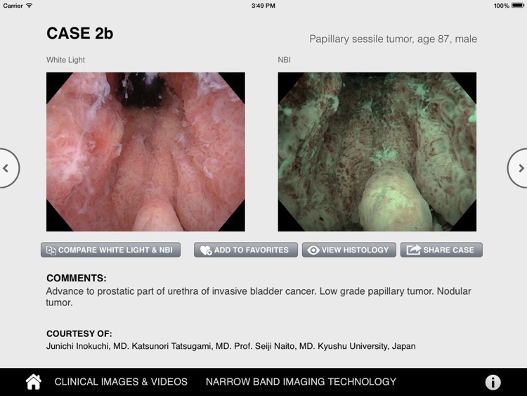
The NBI Atlas for Urology has been developed in collaboration with world-renowned urologists from North America, Asia and Europe. Designed as an educational tool, it allows clinicians to view different image sets in white light and NBI light to advance their understanding of endoscopic imaging using Narrow Band Imaging (NBI) technology. Histology is also included for many cases.
App Screenshots
App Store Description
The NBI Atlas for Urology has been developed in collaboration with world-renowned urologists from North America, Asia and Europe. Designed as an educational tool, it allows clinicians to view different image sets in white light and NBI light to advance their understanding of endoscopic imaging using Narrow Band Imaging (NBI) technology. Histology is also included for many cases.
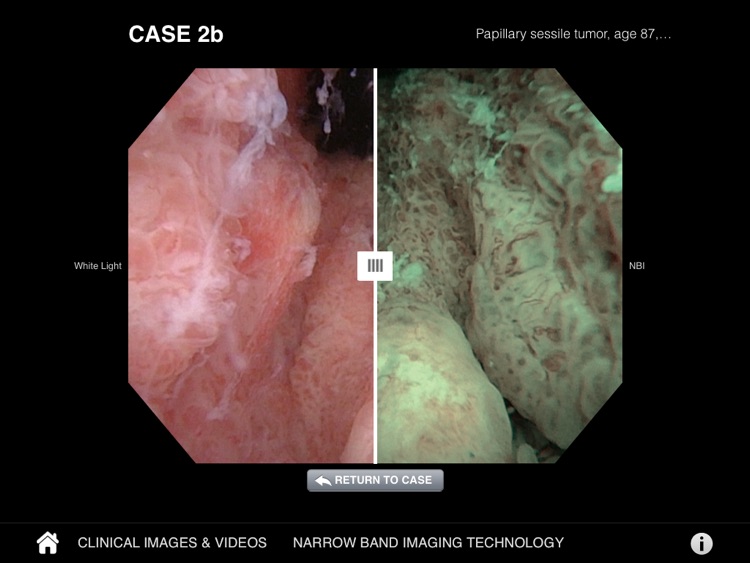
This detailed interactive atlas includes more than 100 high resolution images and emphasizes those conditions for which NBI is considered particularly valuable. Of particular interest is the comparison tool that makes use of a slider handle to show and reveal images in white light vs. NBI.
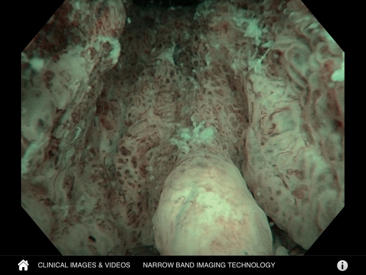
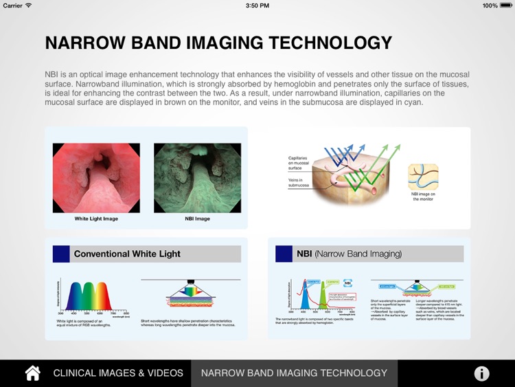
NBI is an optical image enhancement technology that enhances the visibility of vessels and other tissue on the mucosal surface. Narrowband illumination, which is strongly absorbed by hemoglobin and penetrates only the surface of tissues, is ideal for enhancing the contrast between the two. As a result, under narrowband illumination, capillaries on the mucosal surface are displayed in brown on the monitor, and veins in the submucosa are displayed in cyan.
Disclaimer:
AppAdvice does not own this application and only provides images and links contained in the iTunes Search API, to help our users find the best apps to download. If you are the developer of this app and would like your information removed, please send a request to takedown@appadvice.com and your information will be removed.
AppAdvice does not own this application and only provides images and links contained in the iTunes Search API, to help our users find the best apps to download. If you are the developer of this app and would like your information removed, please send a request to takedown@appadvice.com and your information will be removed.