Intramedullary nailing (IMN) is commonly used to address diaphyseal fractures of long bones
IMNailScrewAR


What is it about?
Intramedullary nailing (IMN) is commonly used to address diaphyseal fractures of long bones. Accurate aiming the distal interlocking holes of the nail for insertion of transfixing screws remains a challenging problem in locked intramedullary nailing requiring intraoperative fluoroscopic guidance. The ‘‘free-hand technique’’ is not sufficiently accurate, because it is conducted by repeated trials using two-dimensional fluoroscopic images. Determining the distal hole using fluoroscopy requires even more radiographic exposure. The greatest level of radiation exposure to the patient and operating room staff was recorded during intramedullary nailing that involves distal interlocking. In an attempt to reduce this potentially harmful radiation burden, several authors proposed fluoroscopy-independent techniques, by proximally mounted target systems. Although these systems improve the operation accuracy, they still rely on radiographic images and the costs of configuring these systems are high.

App Screenshots


App Store Description
Intramedullary nailing (IMN) is commonly used to address diaphyseal fractures of long bones. Accurate aiming the distal interlocking holes of the nail for insertion of transfixing screws remains a challenging problem in locked intramedullary nailing requiring intraoperative fluoroscopic guidance. The ‘‘free-hand technique’’ is not sufficiently accurate, because it is conducted by repeated trials using two-dimensional fluoroscopic images. Determining the distal hole using fluoroscopy requires even more radiographic exposure. The greatest level of radiation exposure to the patient and operating room staff was recorded during intramedullary nailing that involves distal interlocking. In an attempt to reduce this potentially harmful radiation burden, several authors proposed fluoroscopy-independent techniques, by proximally mounted target systems. Although these systems improve the operation accuracy, they still rely on radiographic images and the costs of configuring these systems are high.
The app allows orthopedic surgeons unimpeded passage of the drill bit through the distal locking hole of a nail by :
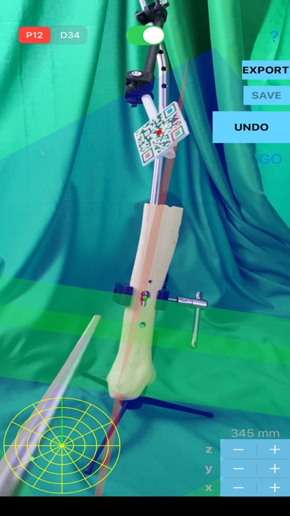
-watching the planes in AR to navigate in real time. After calibration - independent from nail manufacture - three perpendicular planes appear in AR passing with different hazy transparent colours through the distal holes. A passive sensor (PS) is used for calibration and acts also as a dynamic reference guide because which is constantly recognised and tracked and updating the position of all registered points to new one. In case the IM nail and the proximally mounted target PS is placed in different position as a whole array, all registered points are placed in new position respectively in AR. The surgeon now can navigate and accurate identify the direction of the drill bit toward the distal hole of the nail.
-observing a ‘’radar ‘’ screen which is depicted on screen and act as a navigation map during screw placement. A dot appears inside the radar screen reflecting the position of the tip in relation to half distance between two screw opening (in-out). Manipulating the handle of screw driver the dot size and location in “radar” screen change respectively, in real time. The size of the dot in radar screen is also changed according to the distance to insertion point. The dot in radar screen is starting to enlarge when the tip of the screw is approaching the entrance of hole namely the closer the tip of the screw the bigger the dot in the radar screen.
-seeing in real time current location of the tip of the screw, in relation to hole quadrants which are also depicted (left up, left down, right up, right down quadrant )
-observing current distance measured from tip of the screw during insertion - in mm - to the entrance hole. When the tip of the screw is approaching the proximal the entrance of the hole, distance is reduced. Once the tip of the screw attaches the centre of the entrance hole , distance becomes 0 in mm.
seeing on screen three colourful columns representing coronal, transverse, sagittal planes, and angles also measured in real time between the previous mentioned planes and the respective planes of tip of the registered screw - values in normal range are taking green colour, otherwise red- which are continuously printed.
The Augmented Reality targeting App can be a robust guidance solution tool for education and training young fellows or residents in orthopaedic surgery by practicing over a benchmark with saw bone before use in real surgery and also might offer an easy to use, precise, low-cost, less time - consuming, complications and radiation-free, technique, allowing novice and expert surgeons alike to perform easily the distal locking screw placement.
AppAdvice does not own this application and only provides images and links contained in the iTunes Search API, to help our users find the best apps to download. If you are the developer of this app and would like your information removed, please send a request to takedown@appadvice.com and your information will be removed.