You are using an outdated browser. Please
upgrade your browser to improve your experience.
Volume 5 in the Series: The Johns Hopkins Atlases of Pathology

Ovarian Tumor Pathology
by Johns Hopkins Mobile medicine
What is it about?
Volume 5 in the Series: The Johns Hopkins Atlases of Pathology
App Screenshots

App Store Description
Volume 5 in the Series: The Johns Hopkins Atlases of Pathology
AUTHORS: Emanuela F. T. Veras, Ayse Ayhan, Tsui-Lien Mao, Marissa J. White, and Norman J. Barker
SERIES EDITORS: Toby C. Cornish, Norman J. Barker, and Ralph H. Hruban
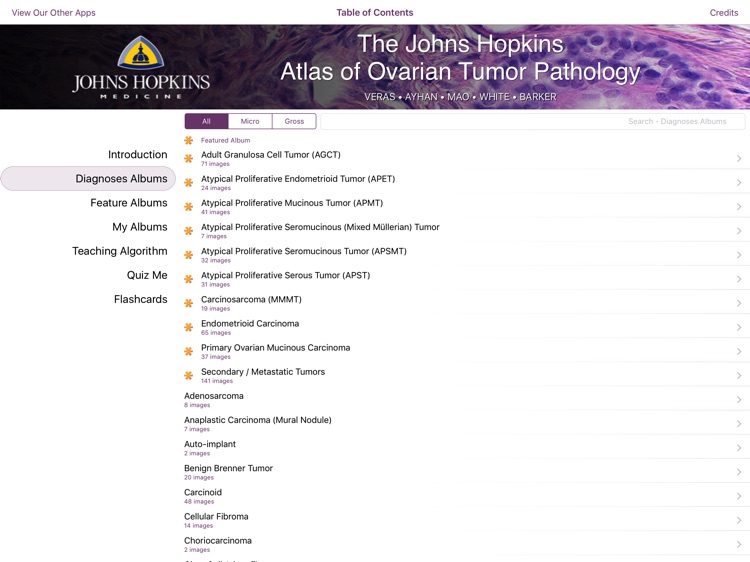
The Johns Hopkins Atlas of Ovarian Tumor Pathology is the fifth teaching app in our series from the Johns Hopkins University Department of Pathology. This app provides an easily accessible atlas with as many as possible images on each specific entity, common and rare documented variations on every neoplasm, along with succinct, explanatory descriptions that would aid the novice in the field as well as the experienced professional to arrive at the correct diagnosis. Although the focus of this work is on neoplastic ovarian lesions, we have also included selected examples of non-neoplastic entities and normal ovarian structures we deem as important to recognize.
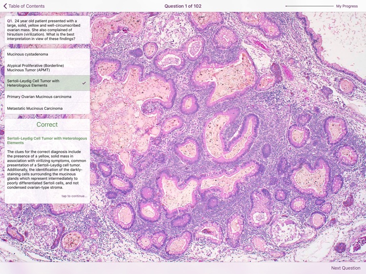
This app follows the format presented in previous atlases of this series. It is composed of four modules: an interactive teaching algorithm, a searchable image atlas, an image-based quiz, and a flash card module.
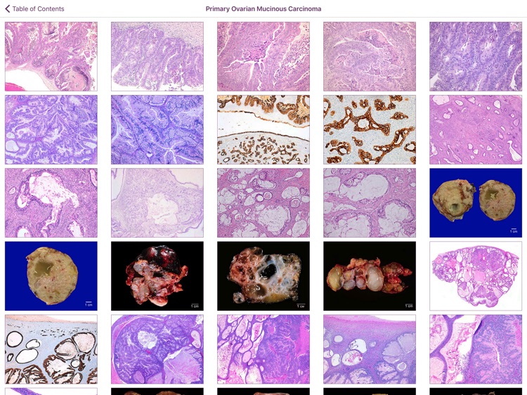
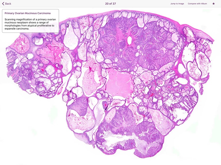
The image atlas contains over 1600 high resolution color images with expertly-authored captions. These microscopic, macroscopic and radiographic images can be viewed together or separately, and two entities can be compared, side-by-side. The atlas can be viewed by diagnosis or by features (these features re-enforce the diagnostic features used in the algorithm), and the atlas can be searched using a key word search. You can create your own albums and add images you select from from the atlas to these albums. The flash cards module allows you to view a randomly generated assortment of images from the image bank and subsequently view the diagnosis and caption by simply “flipping” the card over.
For our teaching algorithm, we present the reader with our general approach to the challenging mucinous tumors of the ovary. This module consists of sequential steps, including approach to the gross (laterality/size)/microscopic characteristics (stratified/non-stratified epithelium) and immunohistochemical profiles of primary and the most common secondary mucinous tumors to the ovary, each step prompting the selection of one out of two presented options, until a final diagnosis is reached.
We welcome your feedback. Please e-mail Dr. Hruban at rhruban@jhmi.edu. If you find an error, please let us know so we can correct it.
Disclaimer:
AppAdvice does not own this application and only provides images and links contained in the iTunes Search API, to help our users find the best apps to download. If you are the developer of this app and would like your information removed, please send a request to takedown@appadvice.com and your information will be removed.
AppAdvice does not own this application and only provides images and links contained in the iTunes Search API, to help our users find the best apps to download. If you are the developer of this app and would like your information removed, please send a request to takedown@appadvice.com and your information will be removed.