Volume 7 in the Series: The Johns Hopkins Atlases of Pathology

Renal Transplant Pathology


What is it about?
Volume 7 in the Series: The Johns Hopkins Atlases of Pathology

App Screenshots


App Store Description
Volume 7 in the Series: The Johns Hopkins Atlases of Pathology
AUTHORS: Serena M. Bagnasco and Lorraine C. Racusen
SERIES EDITORS: Toby C. Cornish, Norman J. Barker, and Ralph H. Hruban
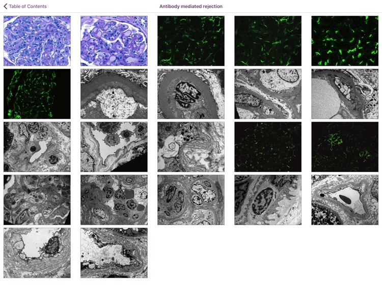
The Johns Hopkins Atlas of Renal Transplant Pathology is the seventh teaching app in our series from the Johns Hopkins University Department of Pathology. This app is designed to teach residents, fellows, and practicing pathologists the basic pathologic lesions in the kidney allograft. The diagnostic entities covered by the Atlas are not limited to rejection but also include recurrent glomerulopathies, infections and other diseases that may affect transplanted kidneys. The diagnostic approach to rejection follows the Banff criteria in the latest version (2017 at the time of this writing). The Banff definitions and rules, Banff scoring system, and Banff classification of rejection are available in text format for easy consultation. The images collected in this Atlas include high resolution examples of light microscopy, immunofluorescence, immunohistochemistry and electron microscopy features of individual diseases. Annotations with comments and references to definition, scoring and classification categories are also included for each image. Three teaching algorithms are provided for navigating through the combination of features leading to diagnosis of specific types of rejection or other conditions unrelated to rejection. These algorithms cover: (1) T-cell mediated rejection, (2) antibody-mediated rejection and (3) other tubular and interstitial pathologic changes. A set of representative images, each with multiple choice quiz can be used for self-test.
We welcome your feedback. Please e-mail Dr. Hruban at rhruban@jhmi.edu. If you find an error, please let us know so we can correct it.
AppAdvice does not own this application and only provides images and links contained in the iTunes Search API, to help our users find the best apps to download. If you are the developer of this app and would like your information removed, please send a request to takedown@appadvice.com and your information will be removed.